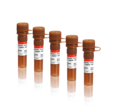
Gel RedSafe Fluorescent Stain - nucleic acid dye for staining DNA or RNA; 10000-fold in water
Suitable to stain dsDNA, ssDNA and RNA, as an alternative to Ethidium Bromide, GelRed or VIVA Sybrgreen Nucleic Acid Stain


Description:
Gel RedSafe is a highly sensitive, non-mutagenic, ultra-safe and ultra-stable fluorescent nucleic acid gel staining reagent, which can replace ethidium bromide EtBr, EB or GelRed from Biotium. Gel RedSafe is higher sensitivity than EtBr without decolorization. Gel RedSafe has the same spectral characteristics as EtBr, and replaces EtBr without changing the imaging system.
Gel RedSafe fluorescent Stain emits red fluorescence when bound to dsDNA, ssDNA, and RNA and replaces the need for the mutagenic ethidium bromide, commonly used in gels staining procedures for visualization of nucleic acids. This product can stain single-stranded DNA and RNA, but is less sensitive to single-stranded DNA or RNA than double-stranded DNA.
DNA visualization after staining with Gel RedSafe fluorescent Stain can be done using conventional gel imaging instruments (UV transilluminators).
The nucleic acid dye is efficiently removed from DNA by gel extraction or ethanol precipitation, and therefore does not interfere with downstream DNA manipulations such as restriction digestion, ligation, PCR, and sequencing.
Gel RedSafe fluorescent Nucleic Acid Safe Gel Stain, 10,000X stock is a concentrated solution. It can be diluted with 5 µl in 50 ml Agarose gel. It can be added directly to the high temperature gel solution.
Features:
- Detection of dsDNA, ssDNA and RNA in gel by pre-staining or post-staining
- Safer alternative to the ethidium bromide staining. Replacement for GelRed from Biotium
- Compatible with various gel imaging instruments
Shipment: @ ambient temperature
Storage: 4-8°C or room temperature. If precipitation is found, please heat the dyes to 45-50°C for 2 min and shake to dissolve, which dose not affect the use.
Final price excl. shipping costs3
- verfügbar / avaílable
- 1 - 3 days for delivery / 1 - 3 Tage Lieferzeit1
How to use Gel RedSafe nucleic acid stain?

Nucleic acid staining. How to make it better?
1. To increase resolution of the DNA bands:
a. Running the gel at a lower voltage for longer period of time
b. Using a wider gel comb
c. Loading less DNA into well
2. To get better separation of bands, adjust the agarose percentage of the gel if the similarly sized bands that bands that are running too close together are loaded. A higher percentage agarose gel will help resolving smaller bands from each other, and a lower percentage gel will help separating larger bands.
Protocol for 1% Agarose Gel
Staining Protocol
A. Pre-cast Protocol (same like as EtBr)
Prepare agarose gel solution using your standard protocol.
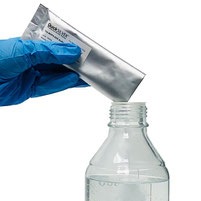
To prepare the gel, add 5 μL of Gel RedSafe nucleic acid dye (10,000 x, water soluble) per 50 mL of agarose gel and mix thoroughly.
Gel RedSafe has excellent thermal stability and can be added directly to the high-temperature gel solution without waiting for the gel solution to cool.
As an alternative: by pre-mixing Gel RedSafe with an electrophoresis buffer containing agarose powder and heating uo.
Note: The pre-cast protocol is not recommended for polyacrylamide gels. Use the post staining protocol for acrylamide gels.
B. Post-staining Protocol
1. Electrophoresis according to conventional methods.
2. Dilute Gel RedSafe Nucleic Acid Dye 10,000 x stock solution approximately 3,300 times to make a 3 x staining solution (for example, add 15 μL of Gel RedSafe Nucleic Acid Dye 10,000 x stock solution to 50 mL of H2O).
3. Carefully place the gel into a suitable container and soak the gel with a sufficient amount of 3 x staining solution. In order to shorten the soaking time, the dyeing solution can be pre-heated to about 70℃, then put into the gel and incubate for 10 min to obtain the ideal effect.
(if not heated, incubate for 30 min in room temperature shaker; if it is acrylamide gel, incubate for 30-60 min and extend with the increase of acrylamide content).
The amount of bubble dyeing dye is large, and the dyeing solution can be reused for about 3 times for a single use. The 3×Gel RedSafe stain solution can be prepared in large quantities and stored at room temperature until used up.
Related products
Deutsche Beschreibung

Datasheet Gel Redsafe Nucleic Stain
Gel RedSafe Stain has the same spectral characteristics as EB, and replaces EB without changing the imaging system.
Material Safety Datasheet
- Safer alternative to the ethidium bromide staining. Replacement for GelRed from Biotium
- Compatible with various gel imaging instruments
References / Protocols / Notes / Recomendations / Tests
Contact
GeneOn GmbH
Carl-Benz-Straße 18
68649 Groß-Rohrheim
Deutschland / Germany
Phone: +49-621-5720 864
Fax: +49-621-5724 462
Shop-Terms
AGB / Business Conditions / Freight Costs / Frachtkosten für Deutschland
GeneOn GmbH
Carl-Benz-Straße 18
68649 Groß-Rohrheim
Deutschland / Germany
Phone: +49-621-5720 864
Fax: +49-621-5724 462
Shop-Terms
AGB / Business Conditions / Freight Costs / Frachtkosten für Deutschland
See Categories
- DNA/RNA Extraction
- Real-time PCR
- RT-qPCR - Master Mixes
- PCR / DNA Amplification
- RT-PCR - Reverse Transcription
- Nucleotides, dNTPs, Primers
- Marker / Gel Staining.
- Enzymes and Fine chemicals
- Fluorescent Dyes / supplements
- DNA/Ladders/Standards
- PCR-96 plates and tools, Tips, Tubes, Filtration
- Devices and Tools
News & Informations
We accept payment through


GeneON GmbH - Germany - Carl-Benz-Straße 18 - 68649 Groß-Rohrheim - www.geneon.net - [email protected]t - Tel: 0621-5720864